
Model: JEM-2100
(Installed: Feb 2011)
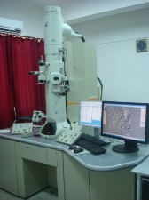
TEM images are formed using electrons transmitted through ultra thin section (50-90nm), thin film or powder. The achievable magnification of the TEM can be from 50X to 1,500,000X and resolution of 0.19 nm depending upon accelerating voltage. The images can be viewed over a fluorescent screen and recorded on a photographic film or a high resolution CCD camera.
Application:
Materials Science, Metallurgy, Biological Sciences, Medical Sciences, Nanotechnology, Ceramics, Pharmaceuticals, Semi-conductors, Polymer Sciences, Drug Delivery Systems, etc.
| Resolution: | 1.9Å to 1.4Å (Lattice) |
| Accelerating Voltage: | 60-200 KV in 50 V steps. |
| Tilt: | ±25° |
| Magnification: | With standard specimen gives x50 to 1,500,000. |
| High Resolution CCD Camera: | 2.672 x 2.672 K |
| Instructions to users: | Biological samples: The specimen should be fixed in 2-3% glutaraldehyde in 0.1M Sodium cacodylate or phosphate buffer (pH 7.2). For biological samples, time gap between sacrifice and immersion in fixative must be minimised. Samples should be trimmed into 1.0- 1.5 mm cube size. Fixation time is 2- 4 hours (depending on tissue type) at 4°C. Wash in 0.1M buffer (3 changes of 15 minutes each) and transport in the same buffer at 4°C. Material Samples: users should write/enquire from the TEM unit for specific instruction according to sample type(s). |
| Accessories: | |
| Biological samples: | Ultra microtome(s), Knife maker |
| Materials Samples: | Energy Dipersive x-ray spectroscopy (EDS), Double Tilt Holder for CBED |
Contact Tel. No. : 0364 2721806; 0364 272 1815; 0364 272 1828.